Advanced Testing to Evaluate Heart Health
Accurate testing is an important part of diagnosing heart disease, evaluating symptoms, and developing the right treatment plan. At Cardiovascular Physicians of Delaware, we offer a wide range of advanced cardiac diagnostic and imaging services using state-of-the-art technology. Whether you're experiencing chest pain, shortness of breath, heart palpitations, or have risk factors for cardiovascular disease, our team can recommend the appropriate testing to better understand your heart health.
Cardiac Imaging & Diagnostic Tests
Echocardiogram (2D)
A 2D Echocardiogram is a non-invasive ultrasound test that creates real-time, two-dimensional images of the heart. It allows doctors to visualize the heart’s chambers, valves, and motion to assess its structure and function. This test is commonly used to evaluate symptoms such as shortness of breath, chest pain, or irregular heartbeats, and to diagnose conditions like heart valve disease, cardiomyopathy, or heart failure. The procedure is safe, painless, and typically takes 30–60 minutes. It provides essential information to help guide treatment and monitor heart health over time.
12 Lead EKG
A 12-Lead EKG (Electrocardiogram) is a quick, non-invasive test that records the electrical activity of the heart from 12 different angles. It helps detect heart rhythm problems, signs of a heart attack, poor blood flow, or structural abnormalities. Small electrodes are placed on the chest, arms, and legs to capture signals, which are displayed as waveforms on a monitor or printout. The test takes only a few minutes and is completely painless. A 12-lead EKG is a valuable tool for diagnosing cardiac conditions and guiding immediate or long-term treatment decisions.
Stress Testing
Stress testing helps evaluate how your heart performs during physical activity or medication-induced stress. These tests can identify reduced blood flow, coronary artery disease, heart rhythm abnormalities, and other cardiovascular conditions while helping your cardiologist determine the most appropriate treatment plan.
Exercise Stress Echocardiogram
An Exercise Stress Echocardiogram is a non-invasive test that evaluates how your heart functions during physical activity. It combines treadmill exercise with ultrasound imaging to assess heart muscle movement and blood flow. The test helps detect blocked arteries, evaluate symptoms like chest pain, and guide treatment decisions. It’s safe, takes under an hour, and provides real-time insight into your heart’s performance under stress.
Dobutamine Stress Echocardiogram
A Dobutamine Stress Echocardiogram is a non-invasive test used when a patient is unable to exercise. Instead of physical activity, the drug dobutamine is administered to make the heart beat faster, simulating the effects of exercise. Ultrasound images are taken to assess how the heart muscle responds to stress. This test helps detect reduced blood flow, evaluate heart function, and diagnose coronary artery disease. It’s safe, typically completed within an hour, and provides important insight into how well the heart performs under stress without the need for physical exertion.
Exercise Stress Cardiolite
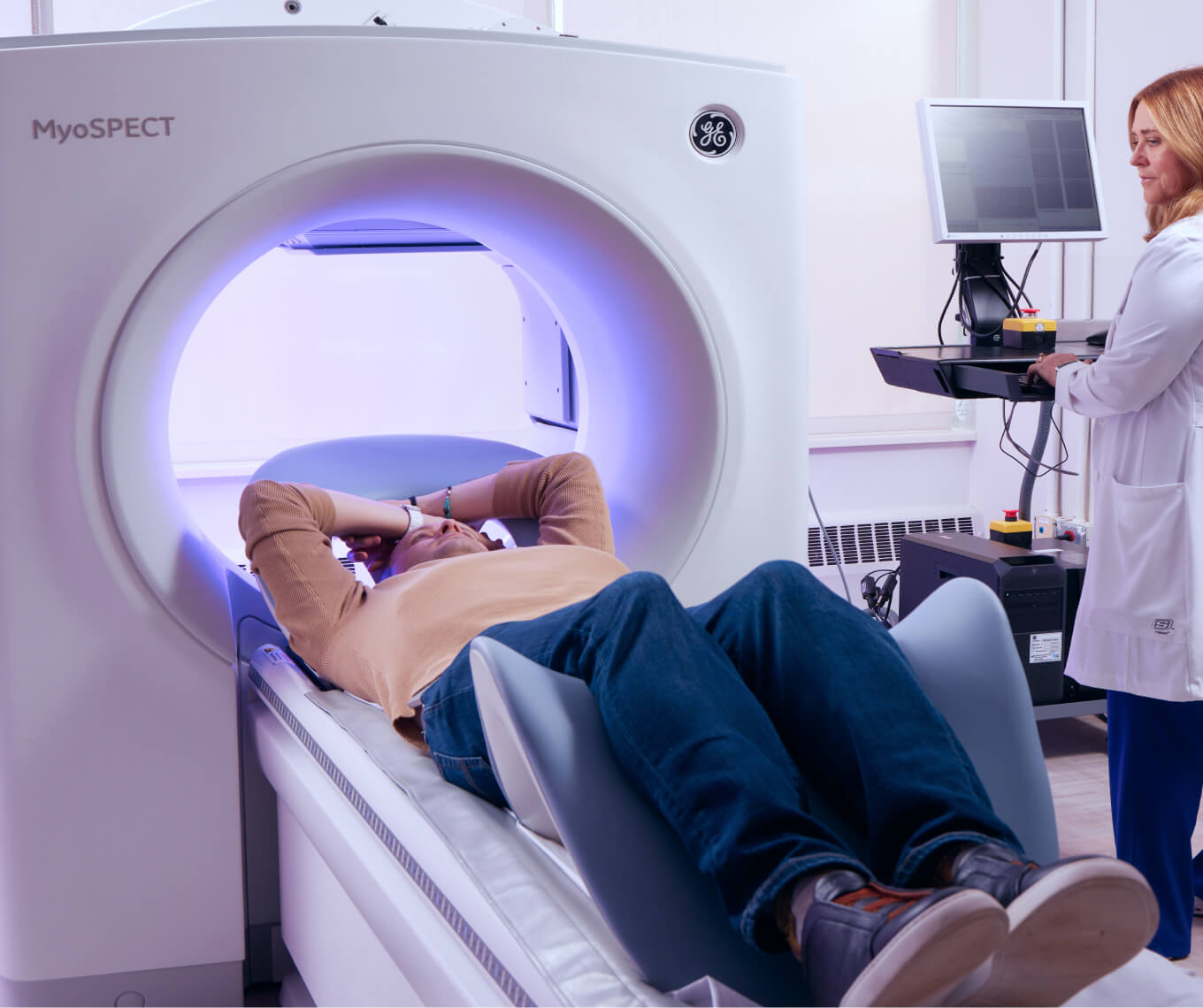
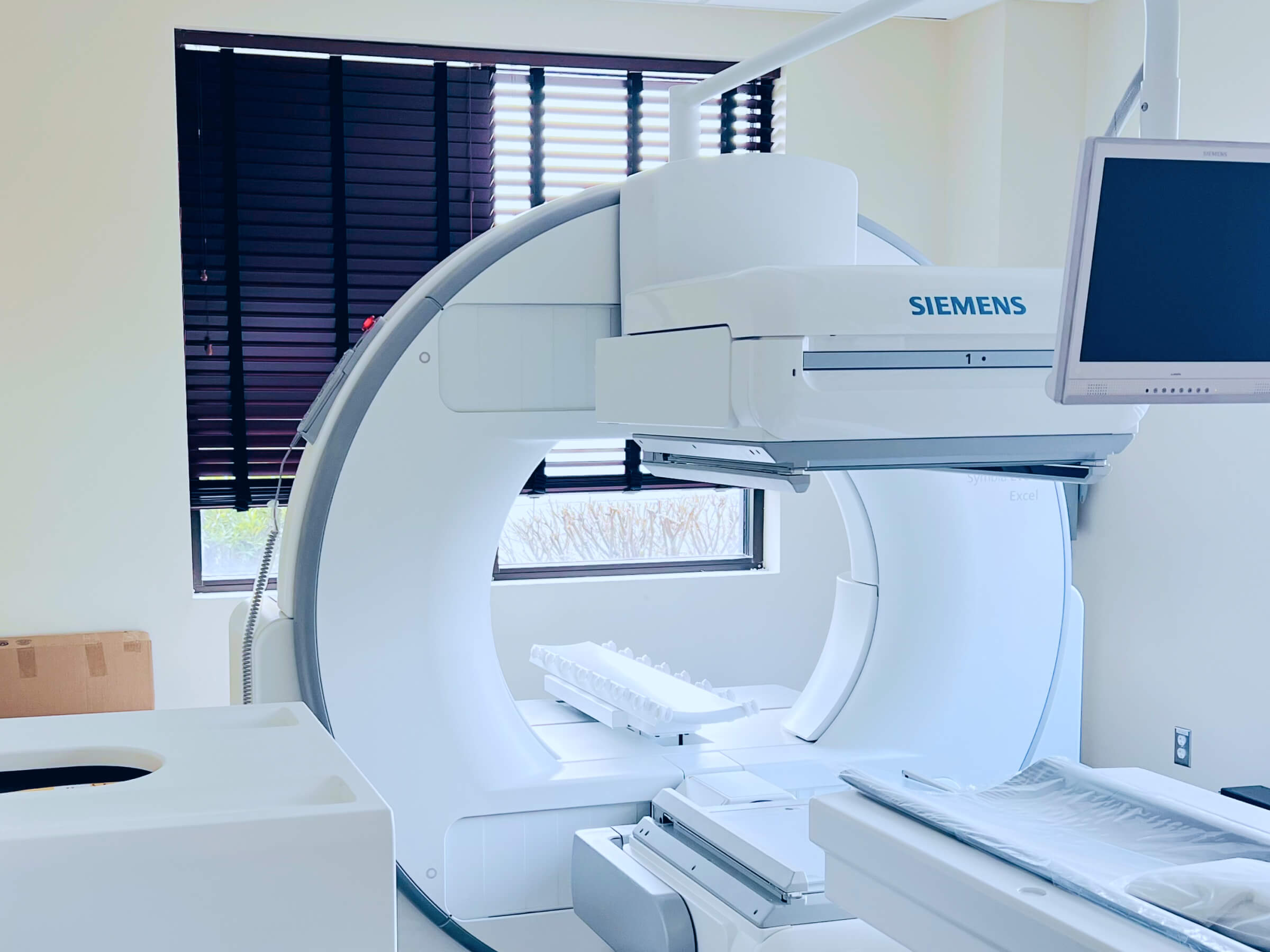
An Exercise Stress Cardiolite test is a nuclear imaging procedure that evaluates blood flow to the heart during rest and exercise. After walking on a treadmill, a small amount of a radioactive tracer (Cardiolite) is injected into the bloodstream. A special camera then captures images showing how well blood reaches different areas of the heart muscle. This test helps detect coronary artery disease, assess damage from prior heart attacks, and guide treatment decisions. It’s safe, effective, and typically completed in a few hours, providing detailed insight into your heart’s blood flow and overall function under stress.
Lexiscan Cardiolite
A Lexiscan Cardiolite test is a nuclear stress test used to evaluate heart blood flow in patients unable to exercise. Instead of using a treadmill, Lexiscan—a medication that increases blood flow to the heart—is administered to mimic the effects of physical activity. A small amount of radioactive tracer (Cardiolite) is then injected, and a special camera captures images of the heart. This test helps detect blocked arteries, assess prior heart damage, and guide treatment. It’s safe, non-invasive, and typically completed within a few hours, providing clear insight into your heart’s function under stress without exercise.
Dobutamine Cardiolite
A Dobutamine Cardiolite stress test is a non-invasive nuclear imaging exam used to evaluate heart function in patients unable to exercise. Dobutamine is given to stimulate the heart, mimicking the effects of physical activity. A small amount of radioactive tracer (Cardiolite) is then injected into the bloodstream. A special camera captures images showing blood flow to the heart muscle at rest and under stress. This test helps detect coronary artery disease, assess damage from previous heart attacks, and guide treatment decisions. It is safe, effective, and usually completed within a few hours.
Advanced Cardiac Imaging
Advanced cardiac imaging provides detailed information about your heart's structure, blood flow, and function. These tests can help your cardiologist diagnose cardiovascular conditions, evaluate your risk of heart disease, and develop a personalized treatment plan based on your individual needs.
Cardiac PET Scan
A Cardiac PET Scan (Positron Emission Tomography) is an advanced, non-invasive imaging test that evaluates blood flow to the heart and detects areas of damaged or scarred heart muscle. It uses a small amount of radioactive tracer and a special camera to produce highly detailed 3D images of the heart. Often used to diagnose coronary artery disease, assess heart function, and evaluate the effects of previous heart attacks, this test offers superior accuracy compared to other imaging methods. Safe and painless, a cardiac PET scan typically takes about 1–2 hours and provides critical insights for guiding treatment.
CT Calcium Score Testing
A CT Calcium Score test is a quick, non-invasive scan that measures calcium buildup within the coronary arteries. Calcium deposits can be an early sign of coronary artery disease, even before symptoms develop. This test helps your cardiologist better understand your risk of future cardiovascular events and determine whether preventive treatments or lifestyle changes may be appropriate. The scan takes only a few minutes, requires no injections or recovery time, and provides valuable information for personalized heart disease prevention.
Heart CT Imaging
Heart CT imaging uses advanced computed tomography (CT) technology to create detailed images of the heart and surrounding blood vessels. Depending on your symptoms and medical history, this test may be used to evaluate coronary artery disease, identify structural abnormalities, or assess blood vessels supplying the heart. Heart CT imaging is non-invasive, highly detailed, and helps your cardiologist make informed decisions about diagnosis and treatment.
Heart MRI Imaging
Heart MRI (Magnetic Resonance Imaging) is a non-invasive imaging test that uses powerful magnets and radio waves to create detailed pictures of the heart and surrounding structures. It can evaluate heart muscle function, detect areas of damage or scarring, assess congenital heart conditions, and provide important information about heart valve disease and cardiomyopathy. Heart MRI offers exceptional image quality without exposing patients to ionizing radiation and can play an important role in diagnosing and managing complex cardiovascular conditions.
When Should You See a Cardiologist for Testing?
Your primary care provider or cardiologist may recommend cardiac testing if you're experiencing symptoms such as chest pain, shortness of breath, heart palpitations, dizziness, or fainting. Diagnostic testing may also be appropriate if you have high blood pressure, high cholesterol, diabetes, a family history of heart disease, or an abnormal EKG. In some cases, testing is recommended before surgery or to monitor an existing cardiovascular condition. Our team will evaluate your symptoms, medical history, and risk factors to determine which tests are most appropriate for you.